ShopDreamUp AI ArtDreamUp
Deviation Actions


Description
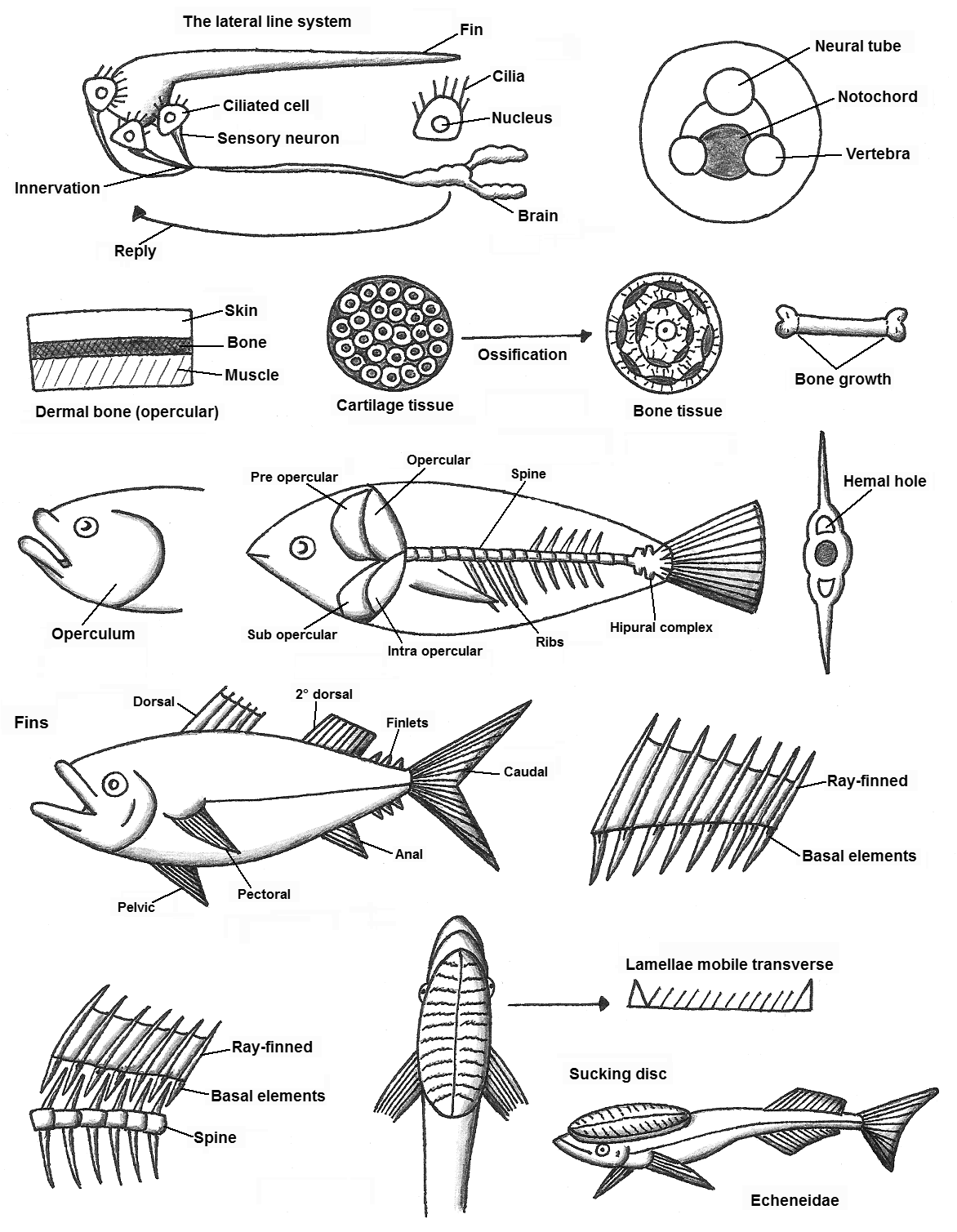
El sistema de la linea lateral en los peces cartilaginosos es un mecanorreceptor, al estimular los nervios que lleva al cerebro y manda respuestas a los músculos y produce el movimiento de las aletas. Este dibujo fue exagerado para exponer sus partes importantes.
Un esquema del cambio de la notocorda en vertebras que son de tejido oseo; en los grupos reptiles, aves y mamíferos.
Diagrama de la estructura de los huesos dermicos. que son planos y anchos, frecuentemente en la cabeza para la protección, también en la respiración (opérculo).
Las células de los tejidos del cartílago y oseo son distinto en forma, estructura y disposición. El tejido del cartílago puede sufrir osificacion para que sea un tejido oseo.
Los huesos en las extremidades en los otros grupos de vertebrados estos crecen desde el nacimiento hasta la vida adulta.
En la cabeza del pez se encuentra el opérculo, que ayuda en el bombeo de agua.
Este operculo esta formado por 4 huesos operculares. En este dibujo se muestra ademas el complejo hipural son las tres ultimas vertebras caudales (las de la cola del pez) de tal manera que se aplanan para formar una “paleta” que utilizan para la locomoción acuática.
Las vertebras en las espinas hay un orificio hemal, en donde entran las arterias.
Dibujo generico de un pez que muestra las aletas, las cuales varían en tamaño, numero y lugar. Todo depende de la especie de pez en el hábitat en donde vive. Las aletas que mas son las pelvicas: inplatacion toraxica, abdominal, en el mentón o puede estar modificada y puede ser una ventosa.
Las aletas están formadas por una membrana y los radios que son huesos. Los elementos basales son huesos con los cuales se fijan los radios.
Las vertebras de la espina no esta unido con los huesos de las aletas.
La rémora un pez de la familia Echeneidae, su primera aleta dorsal esta modificada en un "disco de succión", el cual lleva entre 10 y 28 láminas transversales móviles que les permite aferrarse con fuerza a la piel de otro animal grande.
____________________________________________________________________________________________
The lateral line system in cartilaginous fish is a mechanoreceptor, to stimulate nerves leading to the brain and sends responses to the muscles and causes movement of the fins. This drawing was exaggerated to expose important parts.
A scheme of changing the notochord in vertebrae which are of bone tissue, in the groups reptiles, birds and mammals.
Diagram dermal bone structure. that are flat and wide, often in the head for protection, also in breathing (operculum).
Cells of bone and cartilage tissues are different in shape, structure and arrangement. Cartilage tissue may undergo ossification to be a bone tissue.
The bones in the limbs in other vertebrate groups they grow from birth to adulthood.
At the head of the fish is the cap, which helps in pumping water.
This operculum is formed by 4 opercular bone. This drawing also shows hipural complex are the last three caudal vertebrae (the tail of the fish) so that flatten to form a "palette" used for aquatic locomotion.
The vertebrae in the hemal spines there is a hole where the arteries enter.
Generic drawing of a fish showing the flaps, which vary in size, number and location. It all depends on the fish species in the habitat where they live. The fins are the pelvic more: inplatacion thoracic, abdominal, chin or can be modified and can be a sucker.
The fins are formed by a membrane and that the spokes are bones. Basal elements are bones which are fixed radii.
The vertebrae of the spine is not attached to the bones of the fins.
The drag on a fish Echeneidae family, its first dorsal fin is modified into a "sucking disc," which takes between 10 and 28 mobile transverse lamellae allowing them to cling tightly to the skin of another large animal.
Image size
1172x1509px 190.93 KB
© 2013 - 2024 Maleiva
Comments6
Join the community to add your comment. Already a deviant? Log In
Nice ref!
I actually put this in my references, because no matter where I look to research fish anatomy, there is never a lot of details about the structure of the operculum and intra-operculum anatomy. Overall this is very helpful. Thankyou so much!